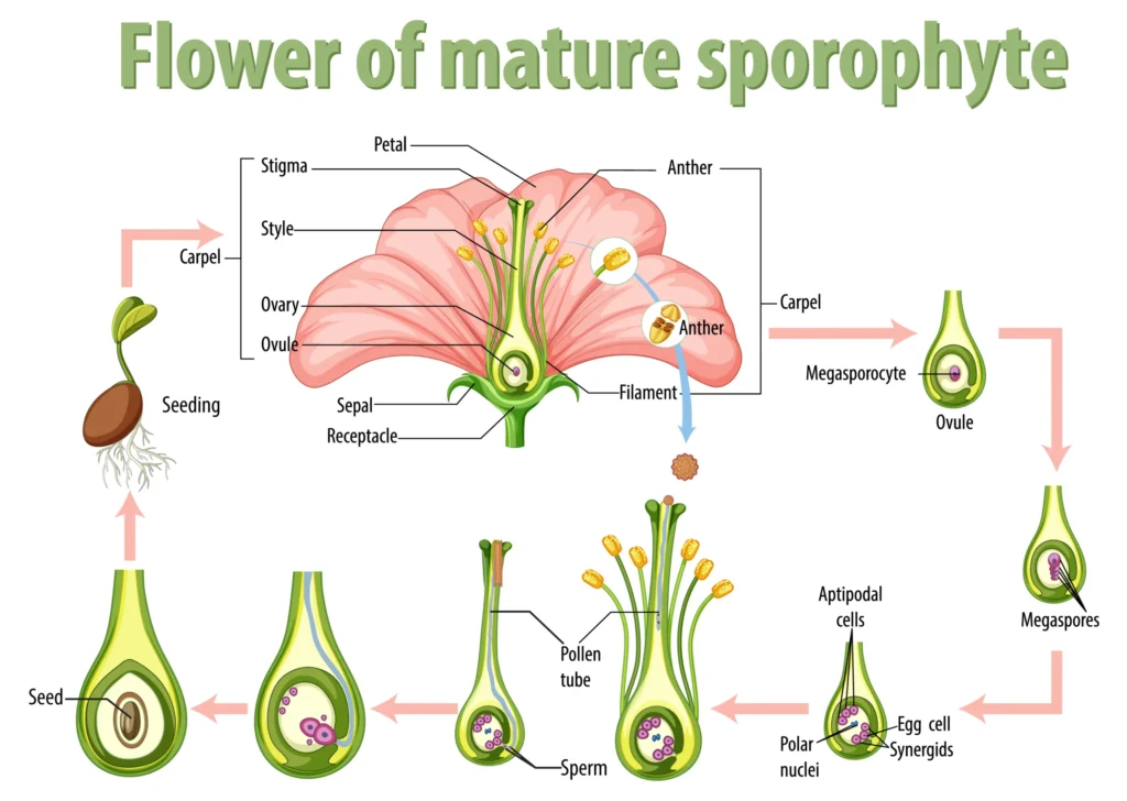
Structure of the Pistil.
The gynoecium represents the female reproductive organ of the flower. The pistil has three parts.
- Stigma
- Stylus
- Ovary
The pollen grain reaches the stigma. The style is a tubular part. The lower, swollen part of the pistil is the ovary.
The gynoecium may contain one or multiple pistils.
1. Monocarpic – Pistils with only one ovary at the base are called monocarpic.
Example – Hibiscus
2. Polycarpic – Pistils with more than one ovary are called polycarpic.
It has two parts.
- Hypocarpic – Pistils with more than one ovary are called syncarpic.
- Dioecious – Multiple pistils that are independent of each other are called dioecious.
The ovary contains a uterine cavity, within which lies the placenta. The megaspores produced in the placenta are called ovules.
Merosporangium (Ovule) – The ovule is a small structure connected to the placenta by a stalk called the micropyle. Each ovule has one or two integuments.
- Outer integument
- Inner integument
The inner integument surrounds the ovule, except for the micropyle. Opposite the micropyle is the chalazal end. Surrounded by the integument is a mass of cells called the nucellus. The endosperm, or female gametophyte, is located in the nucellus. One embryo sac is located within one nucellus. The nucellus provides nutrition to the growing megaspore cell.
Merosporogenesis.
The process of megaspore formation from megaspore mother cells is called megasporogenesis. The nucellus contains a lower layer of cells that divide to form two cells.
- Spore-forming cell
- Parallel cell
This division is mitotic.
The sporogenic cell later functions as the megaspore mother cell. The megaspore mother cell undergoes mitotic division, producing four haploid cells. Three of these cells are destroyed.
Female gametophyte.
One functional cell remains. The process of developing an embryo sac from a single megaspore is called monosporic development.
The functional spore undergoes mitotic division, forming two cells. One of these cells migrates toward the basolateral end and the other toward the micropyle. This forms a 2-nucleated embryo sac. Two cells divide by mitosis into a four-celled 4-nucleated embryo sac, and these four cells divide by mitosis into an eight-celled 8-nucleated embryo sac. After the 8-nucleated stage, the cell wall is formed, forming the embryo sac. Six of the eight nuclei are surrounded by wall cells. The remaining two nuclei are called polar nuclei, located within the central cell.
Structure of the Mature Embryo Sac.
The distribution of cells within the embryo sac is distinctive. Three cells at the micropylar end join together to form the egg apparatus, or aggregate. This consists of two synergids and one egg cell. The synergids play an important role in guiding the pollen tubes. Three more cells are located at the basolateral end, called antipodal cells.
A typical angiosperm embryo sac is 8-nucleated and 7-celled when mature.
")


 in Hindi")


 of a Cell in Hindi.")


 of a Cell.")
